
Ophthalmology Case Study
Equine Vets in Derbyshire & Nottinghamshire

Case Studies
Corneal Ulcer
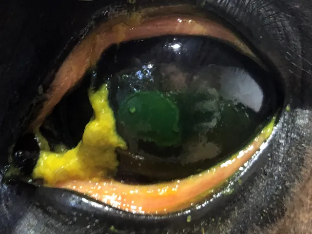
The glassy clear layer at the front of the eye is called the cornea. There are several layers of tissue which form the cornea. These are the epithelium, the stroma and the endothelium (outside to inside). When the outer layer of the cornea (epithelium) is removed, this is termed a ‘corneal ulcer’. There are many inciting causes of a corneal ulcer ranging from direct trauma, e.g. twig or whip injury, to immune-mediated disorders. Corneal ulcers are extremely painful and can lead to inflammation within the eye (Uveitis), which if not controlled, may lead to permanent damage to the eye and unmanageable pain. Please seek advice if your horse has swollen eyelids or appears unable to open the eye to the same degree as the unaffected eye.
The photos below show the ulcerated region of the cornea has been stained with a green dye.

Register your horse or pony with GEM Equine
Register with us today by completing out our online form.


