
Lameness lnvestigations
Equine Vets in Derbyshire & Nottinghamshire

Georgina Parkin
(BVSc., Cert E.P., MRCVS.)
Email: enquiries@gemequine.co.uk
Phone: 07791806970
Lameness In Horses
Lameness is one of the most common problems horses are presented to veterinarians.
How can I tell if my horse is lame?
Lameness is generally best seen in trot. This is a two-time gait and therefore a head nod or hip hike can be better appreciated and attributed to the correct limb than in the other three and four-time gaits unless the lameness is severe in which case the horse may hold the limb abnormally when standing or deviate the limb as he/she moves.
How will the lameness be investigated?
Lameness investigations will be tailored to each individual case. A general approach would be to take a full history, noting the owner’s observations. Then a general assessment of limb conformation, shoeing and stance will be undertaken. This would be combined with a full examination of the limbs by feeling them individually and flexing each joint to assess for abnormal lumps, bumps, thickenings, heat and pain elicited on flexion. The horse would then be trotted up in hand on a flat firm surface to assess for a head nod or hip hike, indicating lameness. Further examination may be carried out on the lunge. Once the offending limb has been identified, further careful palpation of the digital pulses would be carried out, if elevated would tend to indicate inflammation within the foot such as a corn, abscess, bruise or other soft tissue trauma within the foot or pastern. All the accessible tendons and ligaments would be examined for size, consistency and pain on palpation.
Lameness Examination

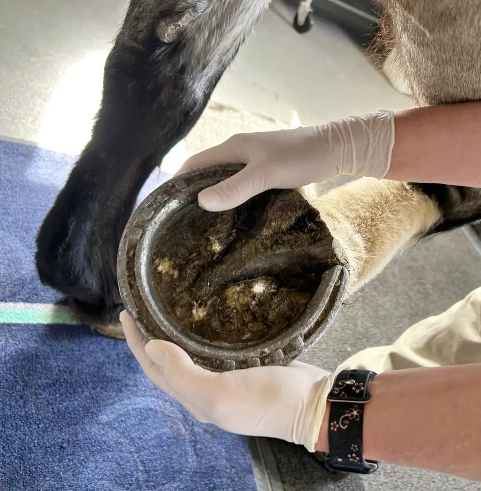
Hoof Examination
The horse hoof would be carefully examined for pain with hoof testers.

Flexion Tests
A series of flexion tests would then be carried out. This is where the joints of the limb are put under stress, and then the horse is trotted up.
This examination is very good at detecting degenerative changes within the joints.

Ultrasound
Gem Equine has a fully portable digital x-ray and ultrasound service available to all clients, which in most cases allows for a swift diagnosis, treatment and management of lower limb lameness. As 95% of lameness originates from the lower limb, most lameness can be managed within the comfort of your own home.
To further localise the site of pain nerve blocks and or blocking of joints might be carried out. A local anaesthetic is used to numb the limb starting with the foot and working up the limb in stages until the horse becomes sound (or much improved). At this stage, when the location of the pain has been identified, imaging techniques might be employed to understand better the full cause of the lameness, its severity, prognosis and therefore guide the vet to give the most effective and appropriate treatment possible. Imaging techniques consist of x-rays (bone examination), ultrasound (soft tissue examination), and more recently, MRI has become increasingly more available to the veterinary market. MRI is especially good at imaging the interior of the foot.


